Single Crystal Notch-Tip Plasticity: Pure Copper
This work appeared in Acta Metallurgica, 44(4),
1996, pp. 1547-1561.
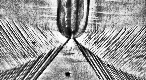 |
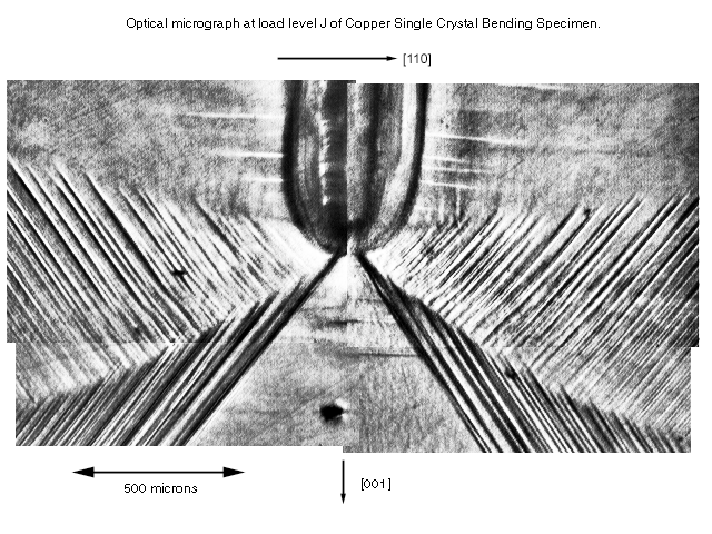
This image shows the surface of the single crystal of copper near
the notch tip after loading and extensive plastic deformation. This pattern of
slip bands in the second and third sectors (counted from ahead of the notch
tip) is quite striking. Compare this pattern with the surface texture that
develops during plastic deformation near a notch tip in a polycrystalline copper specimen. |
|
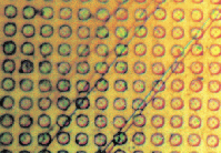
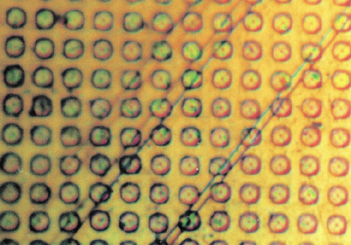
The slip bands on the specimen surface are very thin and the
large amount of shear present in them is visible as offset in the grating dots
the slip lines pass through. |
 |
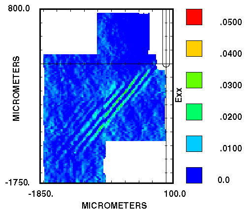
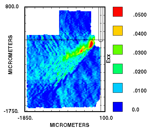
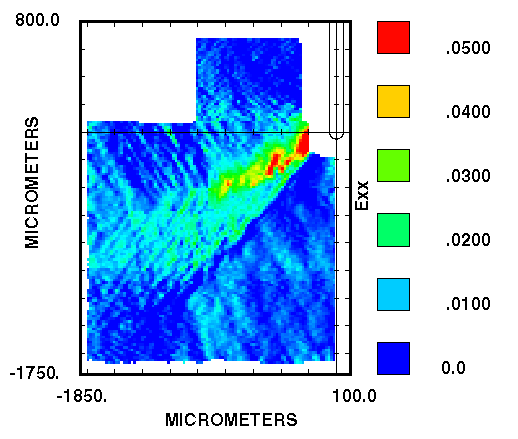
Below are images and movies of the notch tip strains in this as grown
(very soft) copper single crystal at various stages in the loading process. The
orientation of this specimen is identical to the orientation of the
Iron-Silicon crystal discussed in [1]. The X-direction is horizontal and the
Y-direction is vertical in these images and movies.
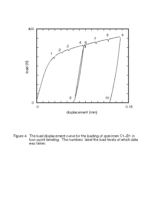
The loading history of the specimen studied here is shown on the
load-displacement plot here.
The time at which data was taken (the load levels referenced below) are
also marked on this figure
Movies of the strain components evolution in false color. The movies
were made by fading between the data in the images below. A solid blue
background was added to avoid the apperance of shifting locations.
Improved Mpeg movies!!
I have come up with a better method of making the mpeg movies of the
strain fields. Instead of fading between images, I now have software that lets
me interpolate between the actual data files before the postscript is produced
that is rendered to produce the images that go into the mpeg file.
 (632 kB) E11 strain component.
-
(599 kB) E22 strain component.
-
(449 kB) E12 strain component.
Old method Mpeg movies
- (1.4 MB) E11 strain
component.
- (1.6 MB) E22 strain
component.
- (1.2 MB) E12 strain
component.
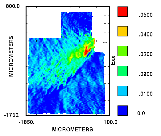
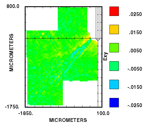
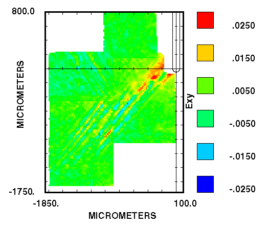
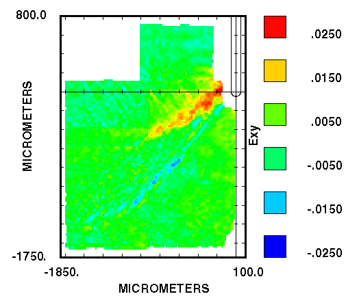
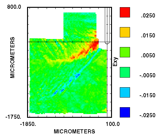
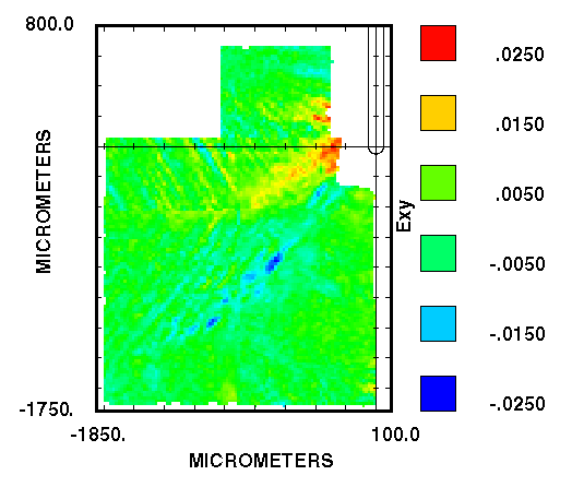
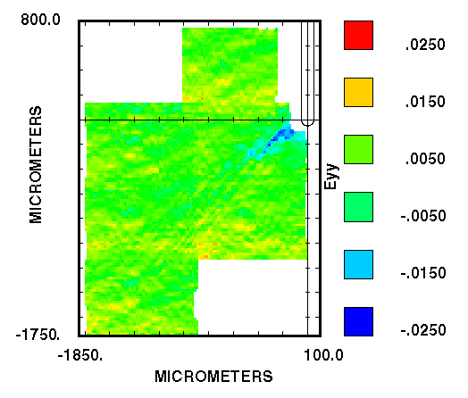
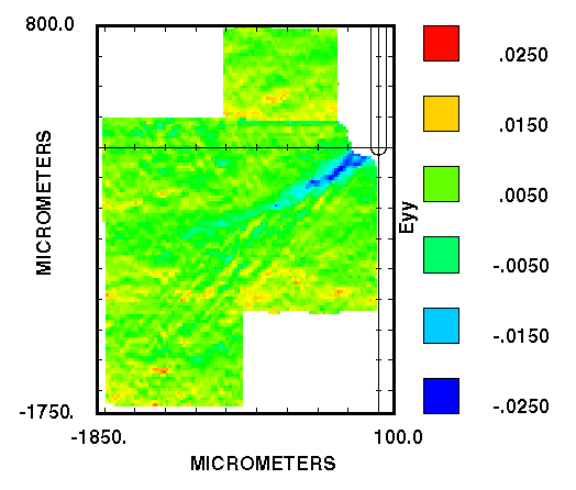
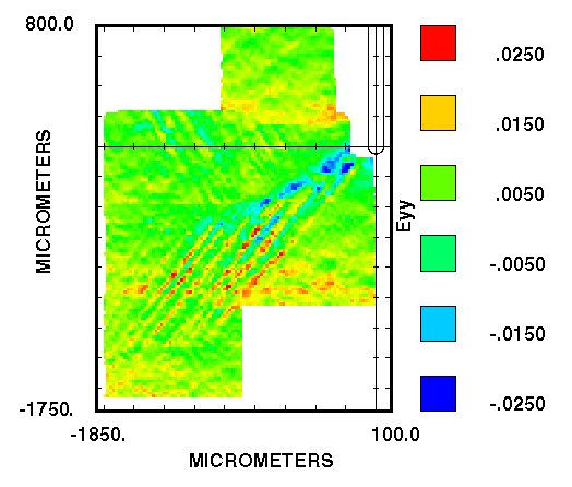
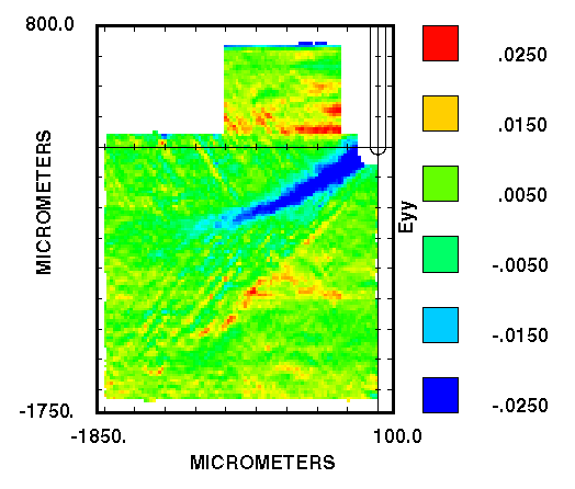
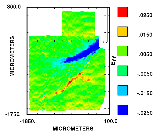
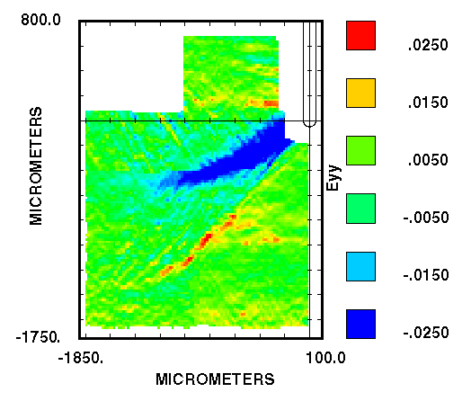
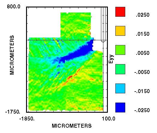
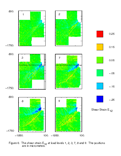
Strain images at the times data was taken, with scales:
- Strain Component E22
- Negative of the Strain Component E12
- Strain Component E11
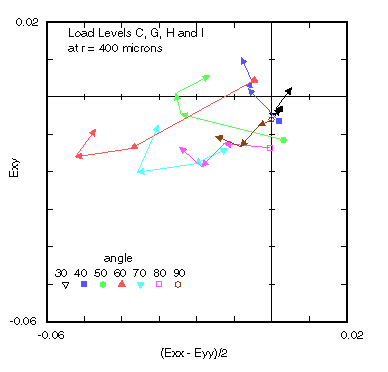
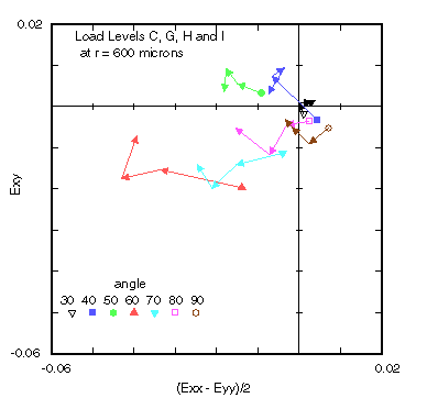
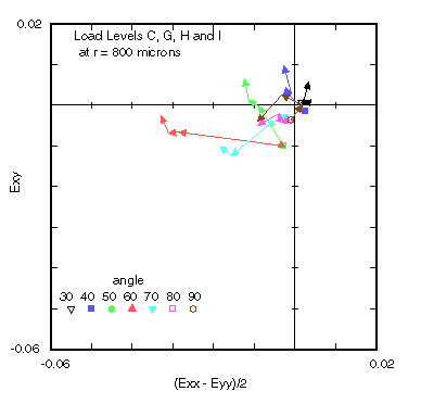
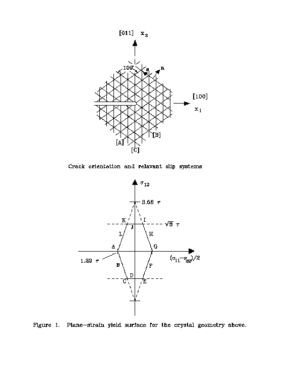
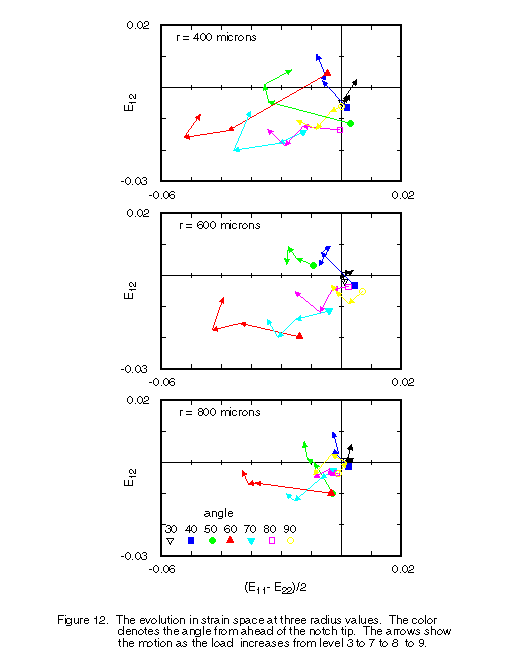
The relavant strain space for this problem is E12 vs (E22 - E11)/2. The
directions in the space can tell us where on the yield surface plasticity
occured. Here are plots in this strain space at 400, 600 and 800 microns
radius. The symbols denote the angle from the crack direction in degrees as
shown. The arrows show the motion of the strain at these locations during
loading from Level 3 to 7 to 8 to 9. These plots show that the loading is
strongly non-proportional.
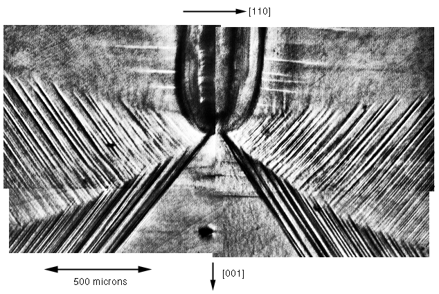
An optical micrograph of the specimen surface taken after unloading
(load level J) is here.
The slip systems in sectors 2 and 3 are clearly visible. This figure also shows
the crystallographic specimen directions.
Figures from the paper:
"An Experimental Study of the Plastic Strain Field near a Notch Tip in a
Copper Single Crystal During Loading," to appear in Acta Metallurgica et
Materialia.
Note that these are low resolution rasterizations of postscript, many
of the figures above are higher resolution.
- Fig 1. The
crystal geometry and yield surface.
- Fig 2. E22
strain component from FE-11.
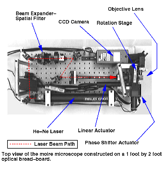
- Fig 3. Over view
of the moire microscope.
- Fig 4.
Load-displacment curve.
- Fig 5. E11 at
load levels 1-3 and 7-9.
- Fig 6. E12 at
load levels 1-3 and 7-9.
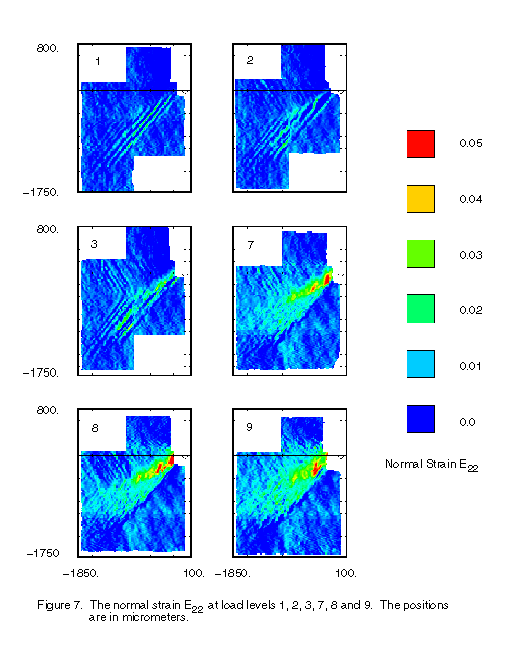
- Fig 7. E22 at
load levels 1-3 and 7-9.
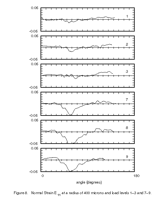
- Fig 8. E11 at r
= 400 microns load levels 1-3 and 7-9.
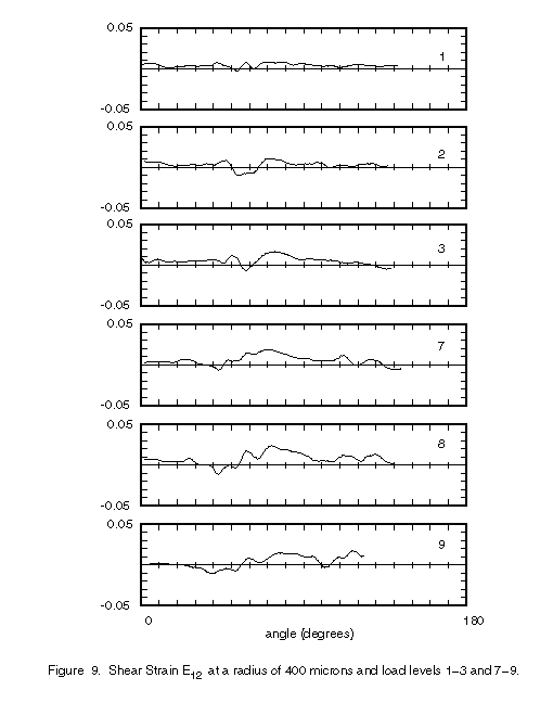
- Fig 9. E12 at r
= 400 microns load levels 1-3 and 7-9.
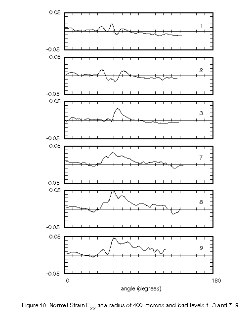
- Fig 10. E22 at
r = 400 microns load levels 1-3 and 7-9.
- Fig 11.
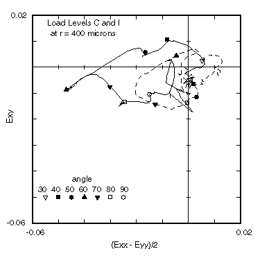
Strain Space at 400 microns load levels 3 and 9.
- Fig 12.
Evolution in strain space r = 400, 600 and 800 microns
- Fig 13.
Optical micrograph of the specimen surface at load level 10.